Ki-67 [SP6] Antibody – 153Eu

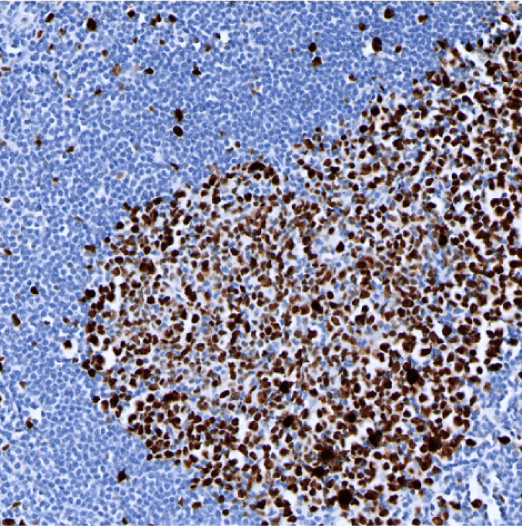
IHC: Ki-67 staining of FFPE human tonsil

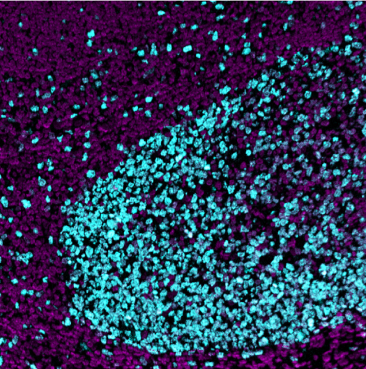
MIBI: Ki-67 staining (cyan) of FFPE human tonsil, costained with dsDNA (magenta)

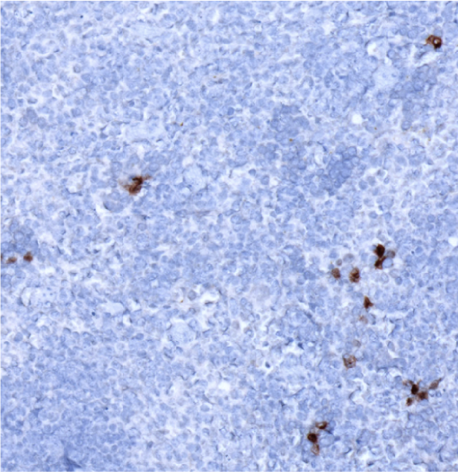
IHC: Ki-67 staining of FFPE mouse spleen

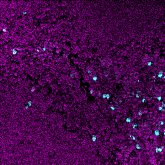
MIBI: Ki-67 staining (cyan) of FFPE mouse spleen, costained with dsDNA (magenta)
Background: Ki-67 is a nuclear protein expressed by proliferating cells (cells in G1, S, G2, and mitosis, absent in the G0 resting phase). The Ki-67 protein has a half-life of only 1–1.5 hours. The Ki-67 proliferation index, or fraction of Ki-67-positive tumor cells, is associated with the clinical course of certain cancers, including carcinomas of the prostate, brain and breast and gastroenteropancreatic neuroendocrine tumors. Antisense oligonucleotides and antibodies targeting Ki-67 have been reported to inhibit cell cycle progression.
Validation: Each lot of conjugated antibody is quality control tested by staining tissue following the MIBI Staining Protocol optimized for the applicable tissue format with subsequent MIBIscope analysis using the appropriate positive and negative tissue field of views.
Recommended Usage: Mouse FFPE: 1 ug/mL dilution. For optimal results, the antibody should be titrated for each desired application.
MIBI technology: Learn more about MIBI™ Technology, a multiplex IHC technology with unmatched sensitivity and true subcellular resolution.
References
Li L.T., Jiang G., Chen Q., Zheng J.N. Ki67 is a promising molecular target in the diagnosis of cancer. Mol Med Rep. 2015;11(3):1566-72.
Rahmanzadeh, R., Rai, P., Celli, J.P., Rizvi, I., Baron-Lühr, B., Gerdes, J., Hasan, T. Ki-67 as a molecular target for therapy in an in vitro three-dimensional model for ovarian cancer. Cancer Res. 2010; 70(22):9234-42.
* Conjugate tested on human tissue and mouse tissue.