LAG3 Antibody – 147Sm
Catalog: 714702
Clone: CAL26
Isotype: Rabbit IgG
Reactivity: Human*
Application: MIBI-FFPE
Storage: Supplied in antibody stabilizer with 0.05% sodium azide. Store at 4°C

IHC: LAG3 staining of FFPE human tonsil

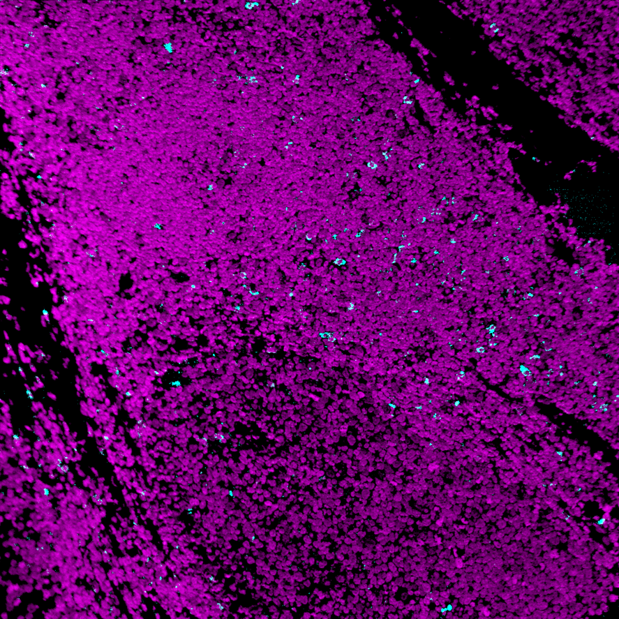
MIBI: LAG3 staining (cyan) of FFPE human tonsil, costained with dsDNA (magenta)

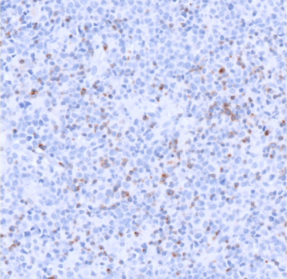
IHC: LAG3 staining of FFPE human high-grade B cell lymphoma

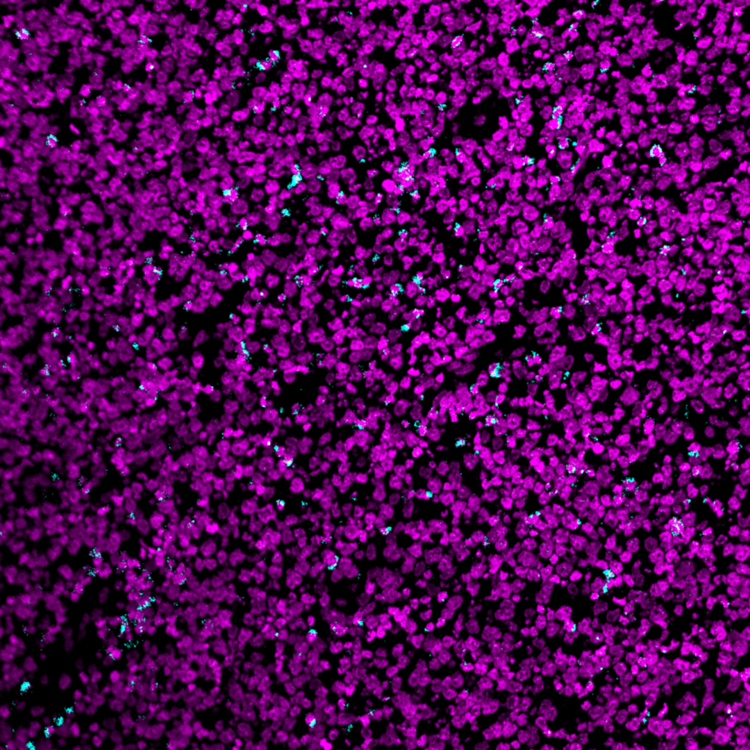
MIBI: LAG3 staining (cyan) of FFPE human high-grade B cell lymphoma, costained with dsDNA (magenta)
Background: Lymphocyte activation gene 3 (LAG-3, CD223) is an immune checkpoint protein that negatively regulates T cells and immune responses. LAG3 is primarily expressed by activated CD4+ T cells, CD8+ T cells, Tregs, and NK cells, where it is activated by MHC Class II molecules, its only known ligand. LAG3 is often co-expressed with PD-1 on the surface of tumor infiltrating lymphocytes, where the two proteins act independently to contribute to tumor-mediated immune suppression. Blockade of LAG3 is a promising strategy for neoplastic intervention.
Validation: Each lot of conjugated antibody is quality control tested by staining tissue following the MIBI Staining Protocol optimized for the applicable tissue format with subsequent MIBIscope analysis of stained tissue microarray using the appropriate positive and negative tissue field of views.
Recommended Usage: Human FFPE: 1 ug/mL dilution. For optimal results, the antibody should be titrated for each desired application.
References
- Seng-Ryong Woo et al. Immune inhibitory molecules LAG-3 and PD-1 synergistically regulate T-cell function to promote tumoral immune escape. Cancer Res. 2012 Feb 15;72(4):917-27.
- Monica V Goldberg & Charles G Drake. LAG-3 in Cancer Immunotherapy. Curr Top Microbiol Immunol. 2011;344:269-78.
* Conjugate tested on human tissue.