MIBIscope™ System
Transforming Multiplexed Tissue Imaging
- Visualize 40+ markers in a single scan
- Comprehensively phenotype immune infiltrate
- Quantify protein expression
- Profile tissue architecture

A revolutionary technology for analysis of the tumor microenvironment
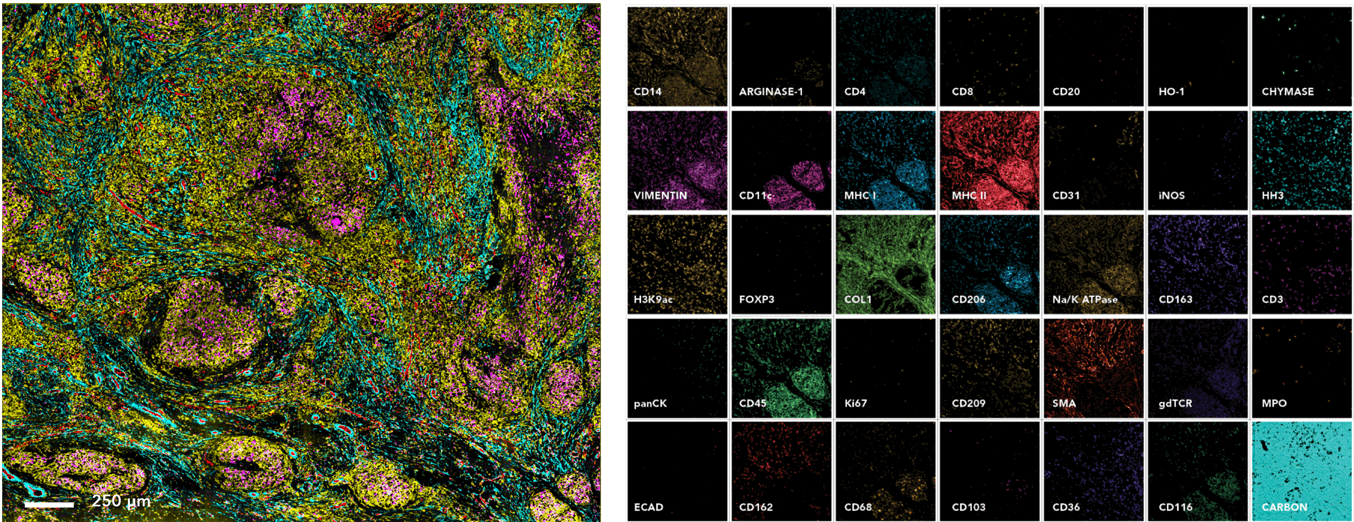
The MIBIscope™ System is a revolutionary imaging platform, enabling comprehensive phenotypic profiling and spatial analysis of the tissue microenvironment. The MIBIscope allows researchers to visualize over 40 markers simultaneously with higher sensitivity, resolution, and throughput than existing methods.

Visualize 40+ Markers in a Single Image
The MIBIscope enables over 40 markers to be visualized simultaneously with single step staining and single step imaging.
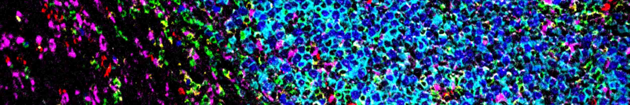
MIBIscope images from a 3mm x 3mm scan of a granulomatous lung section from a Mycoplasm tuberculosis infected patient. The image to the left shows immune infiltrate in the infected tissue (CD45, yellow; CD31, red; SMA, blue; CD68, magenta). Zoomed in single-channel images shown to the right.
High Throughput
Image up to 90 800×800 µm2 ROIs per day
The MIBIscope has the reliability to run 24/7, allowing researchers to conduct studies on large clinical cohorts and image hundreds of samples a week.
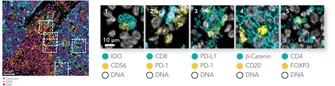
High Resolution
Achieve confocal resolution
The MIBIscope can be adjusted like an optical microscope with resolution settings from 350 nm to 1 µm. At higher magnifications the MIBIscope provides comparable resolution to confocal microscopy.
Data adapted from Keren et al., Science Advances, 2019.
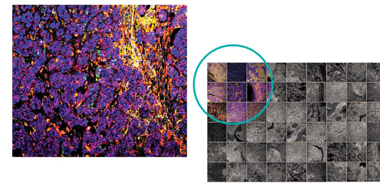
High Sensitivity
Single molecule sensitivity
The MIBIscope uses highly sensitive Secondary Ion Mass Spectrometry, allowing for single molecule detection as demonstrated in Science Advances and other recent publications.
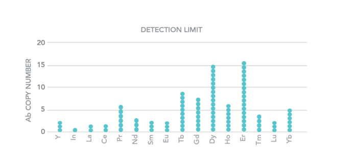
Data adapted from Keren et al., Science Advances, 2019.
SPECIFICATIONS
| Available Biomarker Channels | 40 |
| Pixel Resolution | 400 nm |
| Variable Spot Size | 350 – 600 µm |
| Speed | 400x400 µm2: 9 min 800x800 µm2: 35 min |
| Lower Limit of Detection (number of Ab) | 1 (113In) – 16 (166Er) |
| Dynamic Range | 105 |
| File Type | TIFF |