PD-L1 Antibody – 149Sm

Catalog: 714902 
Clone: E1L3N (PD-L1) & 1D4-C5 (Biotin)
Isotype: Rabbit IgG (PD-L1) & Mouse IgG2a (Biotin)
Reactivity: Human* (PD-L1)
Application: MIBI-FFPE
Storage: Supplied in antibody stabilizer with 0.05% sodium azide. Store at 4°C
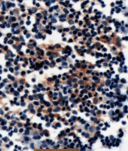
PDL1-IHC-staining-FFPE-human-diffuse-large-B-cell-lymphoma

IHC: PD-L1 antibody staining of FFPE human thymus
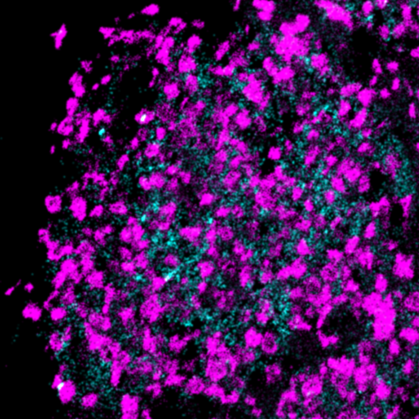
PDL1-Ionpath-MIBI-staining-FFPE-human-diffuse-large-B-cell-lymphoma

MIBI: PD-L1 antibody staining (amplified with anti-Biotin, cyan) of FFPE human thymus, counterstained with dsDNA (magenta)
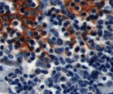
PDL1-IHC-staining-FFPE-human-thymic-SCC

IHC: PD-L1 antibody staining of FFPE human lymphoma
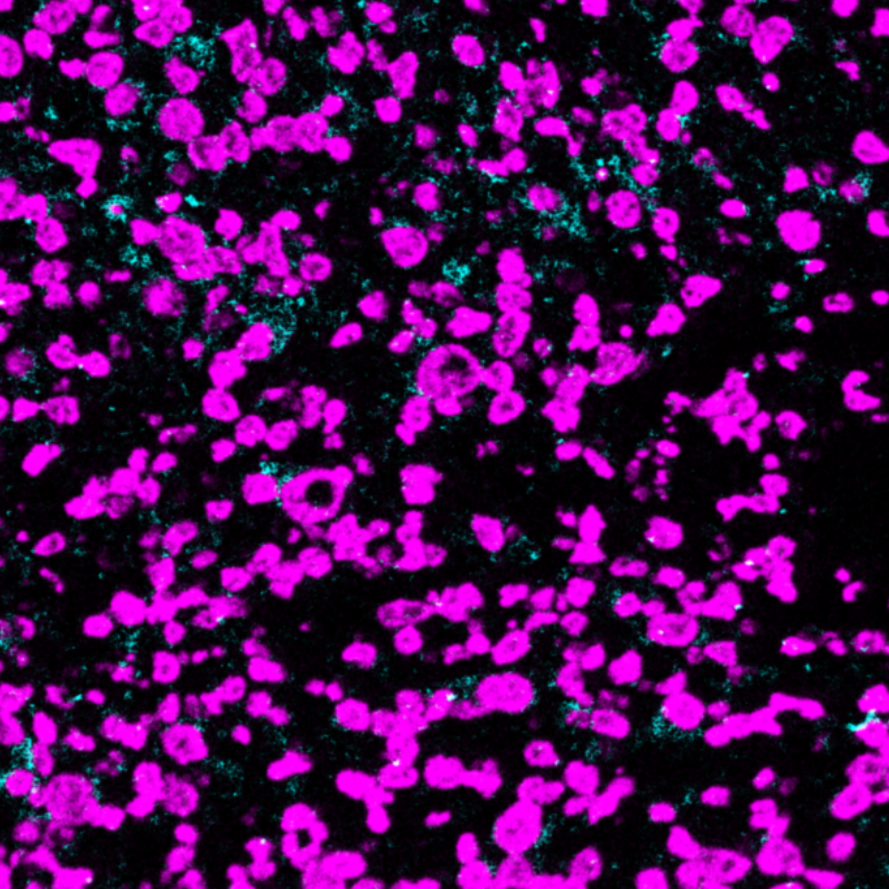
PDL1-Ionpath-MIBI-staining-FFPE-human-thymic-SCC

MIBI: PD-L1 antibody staining (amplified with anti-Biotin, cyan) of FFPE human lymphoma, counterstained with dsDNA (magenta)

Background: Programmed cell death 1 ligand 1 (PD-L1, CD274) binds to PD-1 and inhibits T cell activation. APCs, activated T cells, and tissues including placenta, heart, and lung can express PD-L1. PD-L1 is expressed in cancer as a means for cancerous cells to inhibit immune responses. PD-L1 has been detected for several tumor types including melanoma, lung, ovary, colon, breast, and renal cell carcinomas. PD-L1 expression in cancer is associated with tumor infiltrating lymphocytes, which mediate PD-L1 expression through the release of interferon gamma. Inhibition of the PD-1-PD-L1 axis has been an active area of clinical research with several approved drugs for multiple indications.

Validation: Each lot of conjugated antibody is quality control tested by staining tissue following the MIBI Staining Protocol optimized for the applicable tissue format with subsequent MIBIscope analysis using the appropriate positive and negative tissue field of views. 

MIBI technology: Learn more about MIBI Technology, a multiplex IHC technology with unmatched sensitivity and true subcellular resolution.

References

  • Wimberly, H. et al. PD-L1 Expression Correlates with Tumor-Infiltrating Lymphocytes and Response to Neoadjuvant Chemotherapy in Breast Cancer. Cancer Immunol Res. 2015; 3(4): 326-332.
  • Bindels, S. et al. Regulation of PD-L1 by SIP1 in human epithelial breast tumor cells. Oncogene. 2006; 25:4975–4985.

* Conjugate tested on human tissue.

Contact Us

<p style="text-align: center; margin-top: 2em;">Let us know how we can help you.</p>


    Immuno-oncology/CancerNeuroscienceInfectious DiseaseImmunologyStem CellOther

    255

    Please contact me. I understand that I can choose to unsubscribe at any time by e-mailing marketing@ionpath.com.

    X
    Contact Us

    We are using cookies on our website

    Please confirm, if you accept our tracking cookies. You can also decline the tracking and continue to visit our website without any data sent to third-party services.