MIBIplus
Ionpath’s new service offering takes you from tissue to answers

With MIBIplus, we offer an all-in-one, single-cell spatial proteomic solution that empowers you to detect more than 50 phenotypes in a single scan. Our service can address projects of all sizes, at any frequency and at any stage of your development program so that you can focus on getting the answers that will move your drug program forward.
Our foundational MIBI technology is the cornerstone of our solution, enabling you to maximize the value of your precious tissue samples. It achieves this by detecting all markers in a single scan, preserving spatial architecture. Furthermore, it simultaneously detects high, medium, and low protein expression.
Our dedicated team of technical experts will analyze your tissue and provide initial actionable results in a comprehensive report.
What’s more, this report is entirely customizable, allowing you to iterate and tailor it to your specific research needs. Should you wish to revisit and reanalyze your data, our user-friendly analysis tool makes it as simple as a couple of clicks.
Discover more in your tissue with MIBIplus – your key to unlocking deeper insights and accelerating your research.
MIBIplus deepens tissue understanding, enabling:
- Identify distinct immune cell types and their location in your tissue samples
- Analyze immune activity within your tumor sample
- Evaluate the functional status of immune cells
- Distinguish between responder and non-responder reactions
- Uncover the mechanism of action for immune system reactions in your samples
- Define spatial signatures
- Compare pre- and post-treatment samples
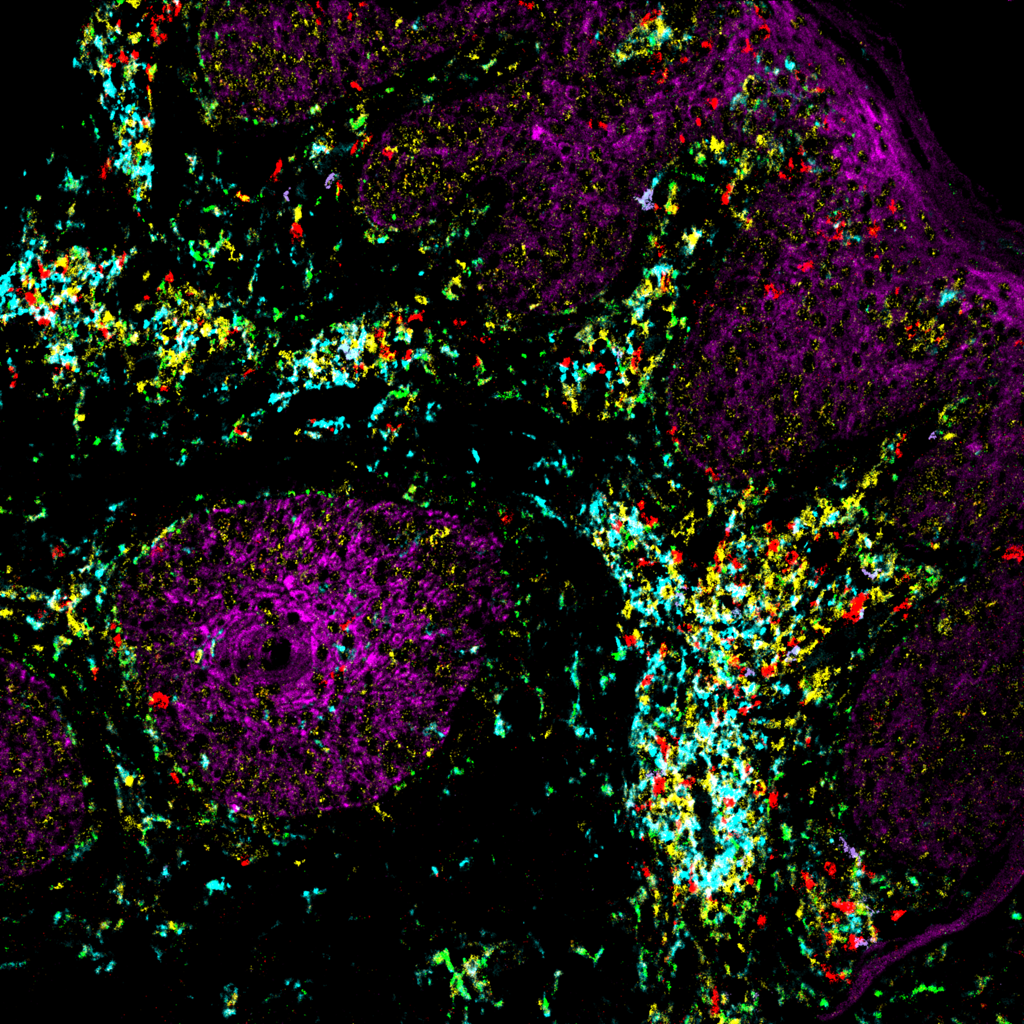
Reveal novel insights into cancer immunobiology with MIBIplus
PROJECT FEATURES
- 50+ cell phenotypes and tissue morphology measured simultaneously from one tissue slide
- Standard, comprehensive spatial analysis report
- Interactive analysis tool
- 27 marker human immuno-oncology panel + 3 advanced checkpoint markers
- Any FFPE tissue except de-calcified
- Mouse immuno-onocology panel coming soon!
PROJECT DELIVERABLES
- CSV files with single cell segmented and phenotyped dataset
- CSV files containing summarized information about the study
- Automated standard report containing quantified aspects of the study
- Access to MIBIplus Manager where single-cell data can be downloaded, visualized and reanalyzed to automatically regenerate a new analysis report.
Try our MIBIplus Pilot Program!
Explore the advantages of MIBIplus by kicking off a pilot project today.
Pilot project includes:
- 2 samples
- Tissue received to report in 4–6-weeks
- Full access to MIBItracker and MIBIsight, our interactive visualization and analysis tools
- Introductory price
MIBIsight
An interactive analysis tool created to streamline analysis and put it back into the hands of biologists
MIBIsight is our user-friendly analysis tools that eliminates the frustration of waiting for your bioinformatics experts to interpret vast datasets. We understand that complex spatial data can often lead to more questions than answers. That is why we’ve designed our interactive analysis tool to quickly get you to results. Our standard reports provide you with an overview of identified cell types, but it doesn’t end there. With just a few clicks, you can navigate and customize your reports and cell classification thresholds, ensuring you have control over your data interpretation. Say goodbye to overwhelming amounts of data and embrace the simplicity of transforming spatial data into clear, straightforward figures that highlight biological differences in your experimental samples. MIBIsight puts the power of comprehensive, actionable insights in your hands, allowing you to receive answers to your specific questions in a matter of weeks, not months.
MIBIplus Service Overview
Unlock Simplified Solutions for Complex Spatial Research
Our dedicated team of technical experts is here to swiftly deliver meaningful readouts from your tissue samples. MIBIplus offers scalability to suit your needs, whether it’s a small-scale project or a large cohort study, all effortlessly managed and tracked through our user-friendly online MIBIplus Manager application. This tool enables simple ordering for new samples as needed, keeps you informed with order status tracking, facilitates seamless communication with our expert Ionpath team, and provides easy access to images and reports. Our service offering is like having your own analytical team, available when you need it, without the hassle or expense of complex instruments. With Ionpath, you can shift your focus to what truly matters – finding answers to your biological questions.
Staining
Our team of MIBI experts receives your tissues that are sectioned on slides, stains them, and identifies the ROIs to be analyzed. Working with you, we verify the selection.

Imaging
On MIBIscope, we simultaneously detect over 50+ cell types in a single scan. The locations of proteins as well as other markers are detected by the instrument and construct a spatial image of proteins without the tissue.

Analysis
Further analysis, takes the raw MIBIscope image to a cell segmented, phenotyped image that can be analyzed and reanalyzed to create a spatial proteomic report without the need for a bioinformatics team.