β-Tubulin Antibody – 113In
Catalog: 711301 
Clone: D3U1W
Isotype: Mouse IgG2b
Reactivity: Human*, Mouse*, Rat, Hamster, Monkey
Application: MIBI-FFPE
Storage: Supplied in antibody stabilizer with 0.05% sodium azide. Store at 4°C

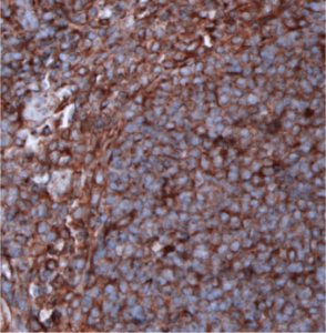
β-tubulin-IHC-staining-FFPE-human-tonsil
IHC: β-tubulin staining of FFPE human tonsil
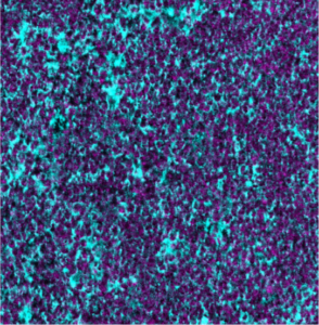
β-tubulin-Ionpath-MIBI-staining-FFPE-human-tonsil
MIBI: β-tubulin staining (cyan) of FFPE human tonsil, counterstained with dsDNA (magenta)
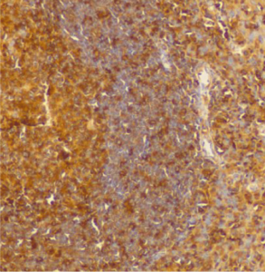
β-tubulin-IHC-staining-FFPE-mouse-spleen
IHC: β-tubulin staining of FFPE mouse spleen
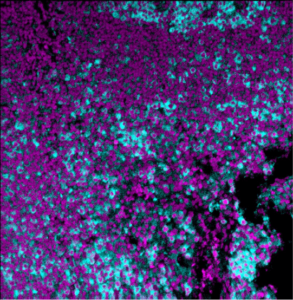
β-tubulin-Ionpath-MIBI-staining-FFPE-mouse-spleen
MIBI: β-tubulin staining (cyan) of FFPE mouse spleen, counterstained with dsDNA (magenta)
Background: The cytoskeleton is composed of microfilaments, intermediate filaments, and microtubules. β-tubulin, together with α-tubulin, forms heterodimers that serve as the building blocks for microtubules. The structure of microtubules is dynamically regulated in processes related to cell movement, cytoplasmic transport, and chromosome alignment during meiosis/mitosis. β-tubulin is broadly expressed in eukaryotic cells. β-tubulin is the target of several anti-tubulin agents used in the treatment of cancer.

Validation: Each lot of conjugated antibody is quality control tested by staining tissue following the MIBI Staining Protocol optimized for the applicable tissue format with subsequent MIBIscope analysis using the appropriate positive and negative tissue field of views. These results are pathologist verified.

Recommended Usage: Human and Mouse FFPE: 1:100 dilution.
For optimal results, the antibody should be titrated for each desired application.

MIBI technology: Learn more about MIBI Technology, a multiplex IHC technology with unmatched sensitivity and true subcellular resolution.

References
Silvia Mihaela Illie et al. Potential clinically useful prognostic biomarkers in triple-negative breast cancer: preliminary results of a retrospective analysis. Breast Cancer (Dove Med Press). 2018; 10: 177–194.

Janke, C. The tubulin code: Molecular components, readout mechanisms, and functions. J Cell Biol. 2014; 206 (4) 461-472.

* Conjugate tested on human and mouse tissue.

Contact Us

<p style="text-align: center; margin-top: 2em;">Let us know how we can help you.</p>


    Immuno-oncology/CancerNeuroscienceInfectious DiseaseImmunologyStem CellOther

    255

    Please contact me. I understand that I can choose to unsubscribe at any time by e-mailing marketing@ionpath.com.

    X
    Contact Us

    We are using cookies on our website

    Please confirm, if you accept our tracking cookies. You can also decline the tracking and continue to visit our website without any data sent to third-party services.