Webinars
Featured Webinar
In this insightful session, we will explore two topics:
Neoadjuvant CD40 Agonism Remodels the Tumor Immune Microenvironment in Locally Advanced Esophageal/Gastroesophageal Junction Cancer
- How MIBI technology complements our understanding of the immune remodeling effects induced by sotigalimab in the tumor microenvironment.
- The mechanisms behind the induction of pathologic complete response rates.
- How sotigalimab enhances antigen presentation and activates cytotoxic T cells.
- Explore the potential implications for improved clinical outcomes in esophageal cancer patients.
MIBIsight: Gaining Insights from Your MIBI Data
- Learn how to harness the power of MIBIsight software for deciphering complex spatial proteomic data.
- Discover how to identify unique signatures and spatial relationships within tissue regions.
- Explore our intuitive tools for adjusting cell classifications and generating comprehensive reports.

Past Webinars
Subcellular Proteomic Imaging Using Multiplexed Ion Beam Imaging (MIBI)
Dr. Jay Tarolli, Ionpath’s Director of Product Development, discusses our MIBI technology, which can detect over 50 cell phenotypes in a single scan.
- Explore how the scientific community applies MIBI to address questions about cancers and diseases
- Dive into the advantages of our time-of-flight secondary ion mass spectrometry (ToF-SIMS) based methodology

Spatial Profiling of Metabolic Regulation
Learn how Dr. Felix Hartmann and collaborators used multiplexed ion beam imaging (MIBI™) technology to develop spatial profiles of metabolic regulation in the human tumor microenvironment.
Dr. Hartmann describes an approach that characterizes the metabolic regulome of individual cells together with their phenotypic identity.
- Using MIBI technology with scMEP, Hartmann and his team characteraized single-cell metabolic features of immune cells in colorectal carcinoma together with their phenotypic identity
- Spatial organization of metabloic programs and distinct metabolic states of immune cells within the tumor micronenvironment were revealed

MIBI Webinar Series
Learn how you can perform comprehensive single-cell analysis, with spatial context—all in one image with multiplexed ion beam imaging (MIBI) technology. This three-part webinar series provides an overview, applications, and data analysis capabilities of MIBI. See how MIBI enables unprecedented, comprehensive analysis of the tumor microenvironment.
- Multiplexed Ion Beam Imaging – Applications in Immuno-Oncology, Neuroscience, and Beyond
- Introduction to Multiplexed Ion Beam Imaging (MIBI)
- Data Acquisition and Visualization of Highly Multiplexed Tissue Images

Comprehensive Enumeration of Immune Cells in Solid Tumors
Presented by Michael Angelo, MD, PhD, Assistant Professor of Pathology, Stanford University
Understanding the role of distinct cellular phenotypes in tissue function, development, and pathogenesis requires tools that can rapidly and consistently quantify the expression of multiple proteins while preserving spatial information. Multiplexed Ion Beam Imaging by Time-Of-Flight (MIBI-TOF) detects elements from hydrogen to uranium, permitting simultaneous measurement of up to 42 labeled antibodies along with histochemical stains and native biological elements. We recently used this capability to analyze infiltrating immune cell populations in archival formalin-fixed paraffin embedded (FFPE) tissue sections from 42 triple negative breast cancer patients. Spatial enrichment analysis showed immune mixed and compartmentalized tumors, coinciding with expression of PD1, PD-L1 and IDO in a cell-type and location-specific manner. Ordered immune structures along the tumor-immune border served as a hallmark of compartmentalization and were linked to survival. We are currently working to build upon this initial effort to develop scoring mechanisms for guiding immune therapy drug selection.
Learning Objectives:
- Learn how MIBI technology enables simultaneous imaging of 40+ markers in FFPE tissue.
- Learn how tumor expression and immune composition are interrelated with histological context that correlates with overall survival in TNBC
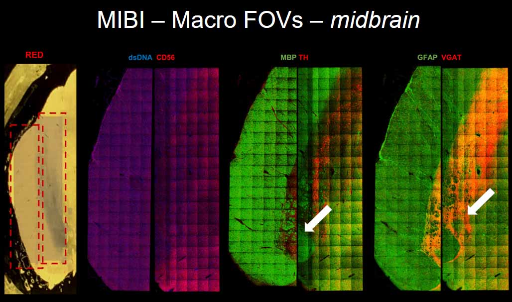
Comprehensive Capture of Human Neuropathology by Multiplexed Ion Beam Imaging (MIBI)
Presented by Sean Bendall, PhD, Assistant Professor of Pathology, Stanford University
Single cell analysis, starting with the earliest low parameter fluorescent experiments, helped define the major cell subsets of human cellular systems as we understand them today. Now, a novel combination of single cell analysis and metal isotopes based mass spectrometry (MIBI) offers a routine examination of 30+ parameters at the nanometer scale, without interference of spectral overlap characteristic of fluorescent reporters. With this platform, we have reached new levels of organizational understanding in human pathobiology – especially when combined with novel single-cell visualization and analysis methods.
Neurodegenerative related disease is estimated to affect five million or more of the ageing American population by 2050. Cognitive care for these individuals is estimated to surpass a trillion dollars a year by the middle of next decade. Accordingly, there is an immediate need for new therapeutic strategies to address this along with identifying unique predictive signatures that correlate with disease mechanism.
During this webinar, we will highlight early applications of this new imaging technology to resolve human neurodegenerative disease and cognitive resilience in retrospective studies of human CNS tissues. This work begins to reveal unappreciated layers of human cellular organization and structure in human systems that can be exploited to understand and perturb human pathobiology.
Learning Objectives:
- Learn the challenges and opportunities for imaging archival CNS tissue
- Learn the principles of Multiplex Ion Beam Imaging (MIBI)
- Learn how MIBI is used to capture multi-scale observations in human neuropathology