CD4 Antibody – 143Nd
Catalog: 714301 Clone: EPR6855 Reactivity: Human*
Storage: CD4 antibody is supplied in antibody stabilizer with 0.05% sodium azide. Store at 4°C
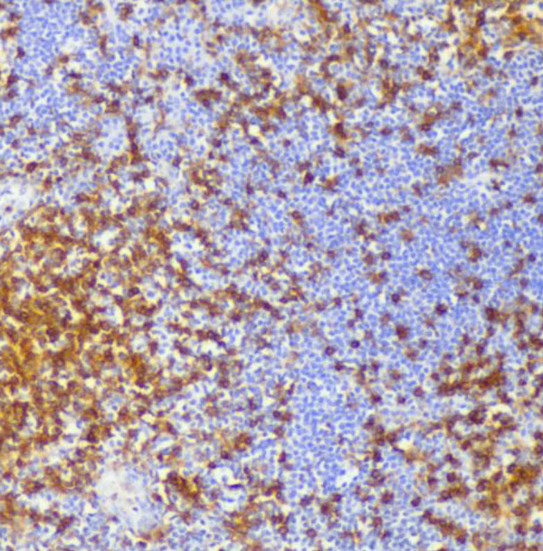
IHC: CD4 antibody staining of FFPE
human tonsil
human tonsil
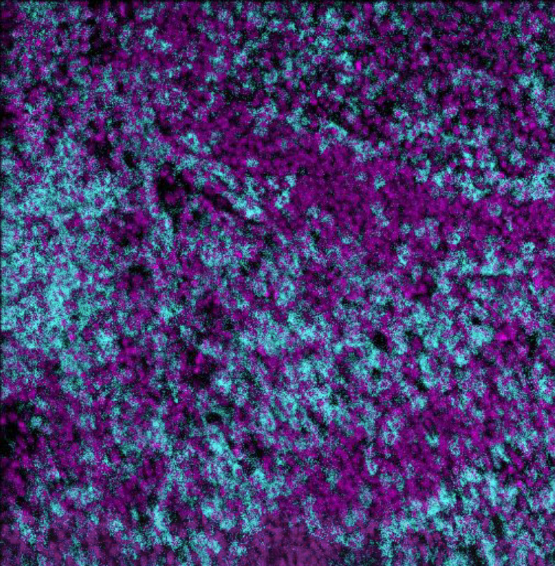
MIBI: CD4 antibody staining (cyan) of FFPE human tonsil, counterstained with dsDNA (magenta)
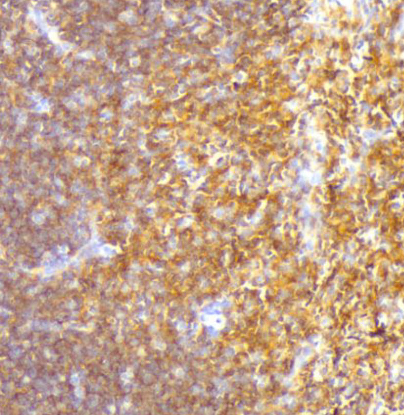
IHC: CD4 antibody staining of FFPE
human thymus
human thymus
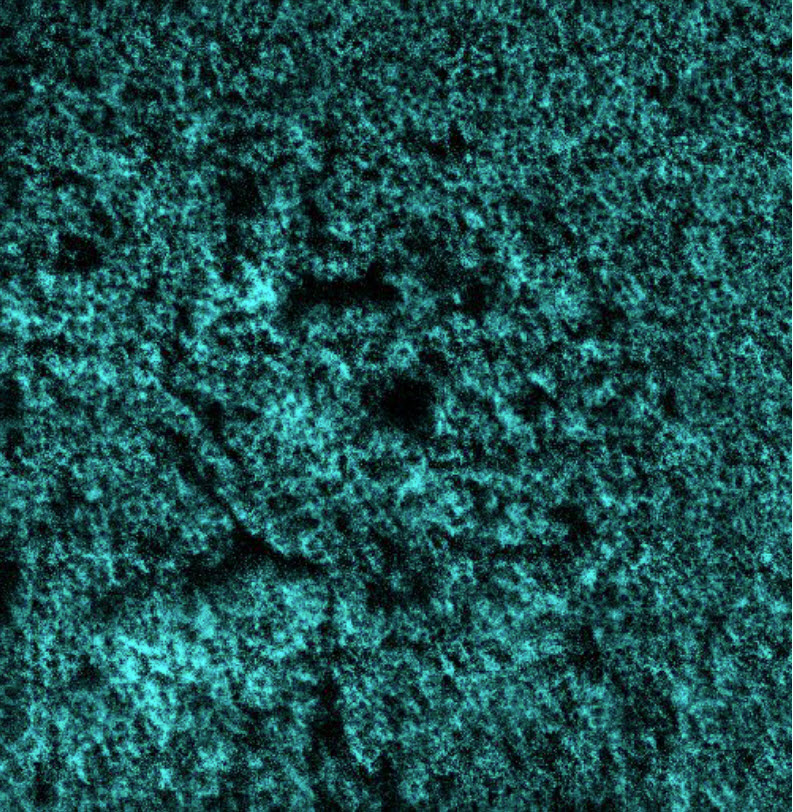
MIBI: CD4 antibody staining (cyan) of FFPE human thymus; dsDNA is not shown.
Background
Background
Background: CD4 is expressed by thymocytes, helper T cells, dendritic cells, type II NKT cells, and monocytes/macrophages. CD4 is a member of the immunoglobulin superfamily and is part of the TCR/CD3 complex, binding to MHC class II molecules and participating in signal transduction through recruitment of tyrosine kinase Lck. CD4 expression is used to identify helper T cells of which there are many different subsets including Th1, Th2, Th9, Th17, regulatory T cell, follicular helper T cell, each contributing to immune function through their unique cytokine profile.
Validation: Each lot of conjugated CD4 antibody is quality control tested by MIBIscope™ analysis of stained tissue microarray using the appropriate positive and negative tissue field of views and are pathologist verified.
Recommended Usage: 1 uL of CD4 antibody per 100 uL staining volume using the MIBI™ Staining Protocol.
For optimal results, antibody should be titrated for each desired application. Suggested starting range is 1:100.
MIBI technology: Learn more about MIBI™ Technology, a multiplex IHC technology with unmatched sensitivity and true subcellular resolution.
References
- Rishi Vishal Luckheeram, Rui Zhou, Asha Devi Verma, and Bing Xia, “CD4+T Cells: Differentiation and Functions,”
Clinical and Developmental Immunology, vol. 2012, Article ID 925135, 12 pages, 2012.
* Conjugate tested on human tissue.