HLA DPDQDR Antibody – 172Yb
Catalog: 717202
Clone: CR3/43
Isotype: Mouse IgG1

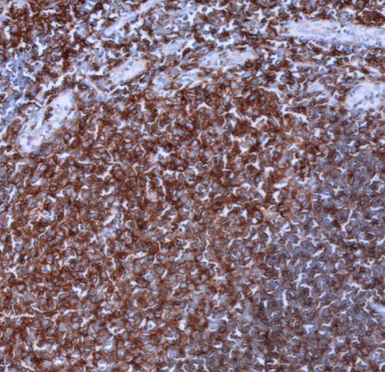
IHC: HLA DPDQDR staining of FFPE human tonsil

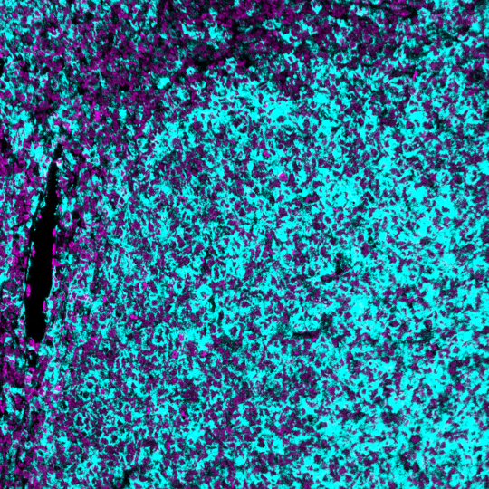
MIBI: HLA DPDQDR staining (cyan) of FFPE human tonsil, costained with dsDNA (magenta)

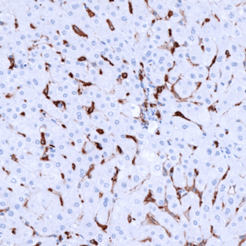
IHC: HLA DPDQDR staining of FFPE human liver

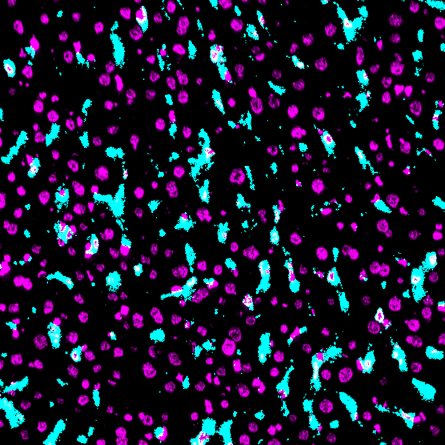
MIBI: HLA DPDQDR staining (cyan) of FFPE human liver, costained with dsDNA (magenta)
Background: MHC class II molecules are transmembrane glycoproteins composed of an alpha chain (36 kDa) and a beta chain (27 kDa). HLA DPDQDR is expressed primarily on antigen presenting cells such as B lymphocytes, monocytes, macrophages, thymic epithelial cells, and activated T lymphocytes. Three loci, DR, DQ, and DP, encode the major expressed products of the human class II region. The human MHC class II molecules bind intracellularly processed peptides and present them to T-helper cells. They therefore have a critical role in the initiation of the immune response.
Validation: Each lot of conjugated antibody is quality control tested by staining tissue following the MIBI Staining Protocol optimized for the applicable tissue format with subsequent MIBIscope analysis using the appropriate positive and negative tissue field of views. These results are pathologist verified.
Recommended Usage: Human FFPE: 1:100 dilution.
For optimal results, the antibody should be titrated for each desired application.
MIBI technology: Learn more about MIBI™ Technology, a multiplex IHC technology with unmatched sensitivity and true subcellular resolution.
References
Thorsby E. Structure and function of HLA molecules. Transplant. Proc. 19, 29.
Daar AS et al. The detailed distribution of MHC class II antigens in normal human organs. Transplantation, 38, 293.
* Conjugate tested on human tissue.