PD-L1 Antibody – 149Sm
Catalog: 714902

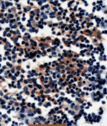
IHC: PD-L1 antibody staining of FFPE human thymus

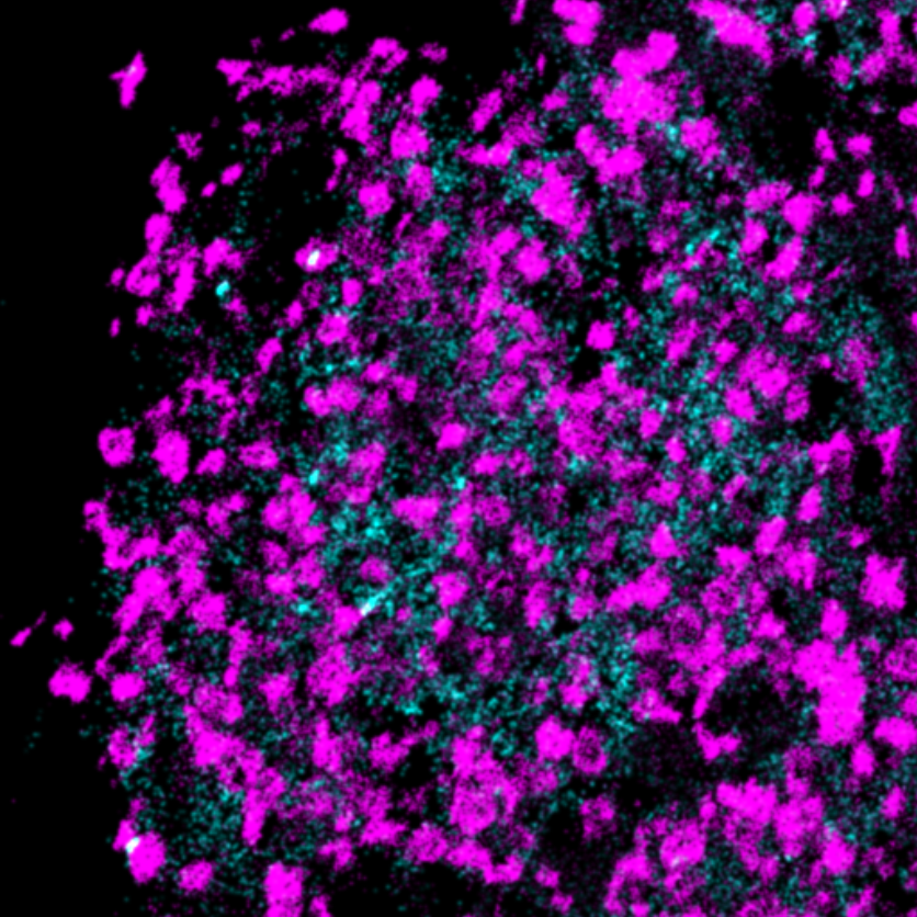
MIBI: PD-L1 antibody staining (amplified with anti-Biotin, cyan) of FFPE human thymus, counterstained with dsDNA (magenta)

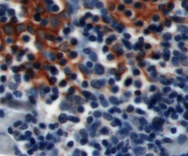
IHC: PD-L1 antibody staining of FFPE human lymphoma

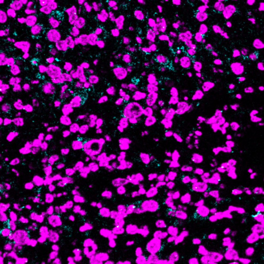
MIBI: PD-L1 antibody staining (amplified with anti-Biotin, cyan) of FFPE human lymphoma, counterstained with dsDNA (magenta)
Background: Programmed cell death 1 ligand 1 (PD-L1, CD274) binds to PD-1 and inhibits T cell activation.
Validation: Each lot of conjugated antibody is quality control tested by staining tissue following the MIBI Staining Protocol optimized for the applicable tissue format with subsequent MIBIscope analysis using the appropriate positive and negative tissue field of views.
MIBI technology: Learn more about MIBI™ Technology, a multiplex IHC technology with unmatched sensitivity and true subcellular resolution.
References
- Wimberly, H. et al. PD-L1 Expression Correlates with Tumor-Infiltrating Lymphocytes and Response to Neoadjuvant Chemotherapy in Breast Cancer. Cancer Immunol Res. 2015; 3(4): 326-332.
- Bindels, S. et al. Regulation of PD-L1 by SIP1 in human epithelial breast tumor cells. Oncogene. 2006; 25:4975–4985.
* Conjugate tested on human tissue.